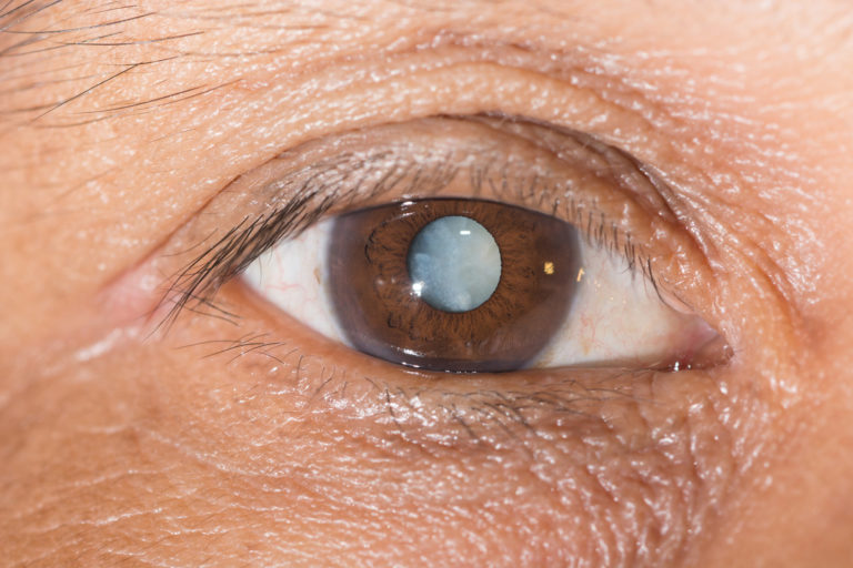
The iLasik ™ platform is indicated for eye treatments under the following conditions: refractive surgeries – Myopia, Hyperopia, Astigmatism and Presbyopia, as well as keratoconus and corneal transplantation.
Myopia, farsightedness, astigmatism, and presbyopia are known as low order aberrations. Personalized treatment with the iLasik TM Platform also corrects high-order aberrations.
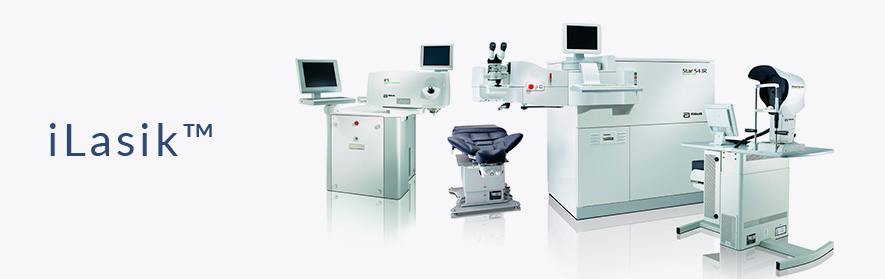
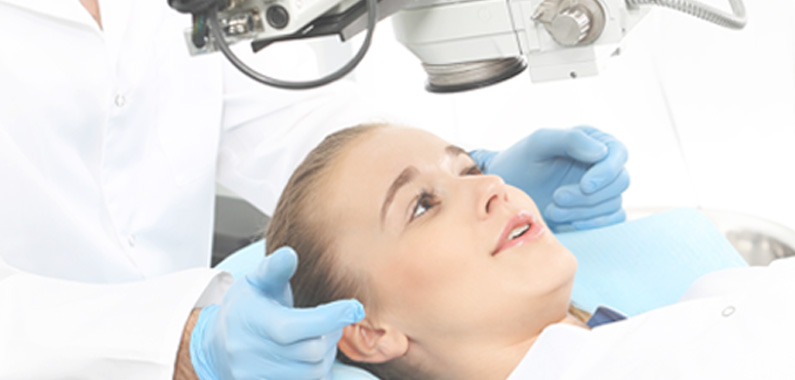
Refractive surgery is a subject already known by the population, but it has particularities that need important clarification. The iLasik ™ procedure represents one of the most advanced technologies for degree surgery and is performed by the Visy Institute, being programmed based on the unique and unique features of your eyes. The three-dimensional mapping of his eyes, and the use of two sophisticated lasers, differentiate 100% laser surgery from other corrective eye-grade corrective surgery that existed previously.
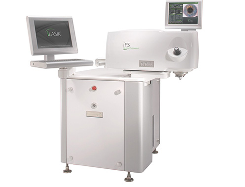
The Visy Institute is a pioneer in this technology in the state of Goiás, and is currently one of the few places in Brazil that has the complete iLasik ™ platform and offers this 100% laser personalized surgery, including FLAP formation for corneal preparation . Our cutting edge in technology and innovation is our trademark, always positioning ourselves at the forefront of the market when it comes to laser refractive surgery and cataract treatment.
There are thousands of people applying to the iLasik ™ procedure. If you wear contact lenses or glasses to see near or far away and want to be free of this discomfort, make an assessment visit to find out if you can benefit from iLasik ™.
Call and bookmark your appointment. +55 (62) 3225-0202 / +55 (62) 99980-2001
It can be affirmed that the treatment for correction of the degree of refraction (Myopia, Hyperopia, Astigmatism and Presbyopia) happens in three simple and fast steps.

1st Stage - Precise Diagnosis
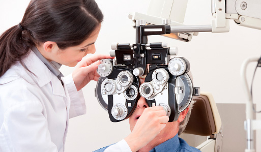
- GREENS: Traditional exam for the diagnosis of the refraction degree (low-order aberrations, hypermetropia, astigmatism and presbyopia). However it is not very specific and measures the degree in intervals of 0.25 D (D = Diopter unit of measure of degree for optical lenses, similar to meter for length). In this way, for example, patients with 5.25 D and another with 5.49 D of myopia will be treated in the same way, with standard measures, ie they will have practically the same glasses, contact lenses or conventional laser treatment.
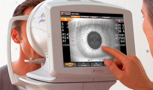
- ABERROMETER: Detailed examination that scans the eye, mapping and generating a digitized three-dimensional (3D) image that identifies Low Order Aberrations (myopia, hyperopia, astigmatism and presbyopia) with a precision of 0.01D intervals (25 times more accurate than traditional diagnosis). In addition to the high order aberrations (coma, trifoil, tetrafoil, spherical aberration, secondary astigmatisms, etc.), these deformities influence the quality of vision, especially at night, as they may present halos around lights, a phenomenon of star explosion (starburst), glare or glare.
The Visy Institute’s WaveScanWaveFront®WaveFronte® AberrMometer, which is part of the iLasik ™ Platform, is undoubtedly a technological breakthrough in the planning of your laser surgery. It accurately identifies the imperfections of each eye, allowing a personalized treatment. The results are automatically reported to the Excimer Laser, which will perform the customized surgery.
In addition to the examination of the degree, some tests are necessary, such as: retinal mapping, ocular pressure measurement, biometry, specular microscopy and ocular tomography.
2st Stage - Preparation of the Cornea
Corneal preparation is directly related to the success of laser eye surgery, whether on efficacy or side effects (irritations, dry eye, infections, etc.).
For greater effectiveness of laser eye surgery, the laser beam has to act accurately. It has to bypass the cornea and reach the stroma to reshape the corneal shape. By changing the curvature, the vision is corrected.
Other techniques for the preparation of the cornea:
PRK – Photo Refractive Keratectomy: Scraping the cornea with a blade to remove tissues from its surface to expose the stroma and allow the laser beam to reach its target in treatment. Disadvantage of this method is slow recovery and there may be pain in some patients. There is a greater risk of infection if the cornea becomes unprotected for a few days until full recovery occurs.
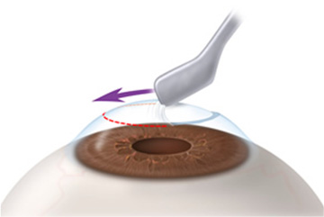
MICROCERATOMY: Mechanical and superficial cut of the cornea with a blade to form the “FLAP” (kind of orange cap) that when raised allows direct and more effective action of the laser. It is not very precise and produces uneven edges and with different thicknesses 120 microns to 180 in their length.
Usually these techniques of preparation of the cornea for the laser surgery irritate more, as they are more aggressive and promote a shock or trauma in the region. Therefore, they require longer post-surgical recovery time, offer greater chances of infection and side effects such as infections, dry eye, flap changes and others.
The American military has banned the flap with a blade, that is, with the microkeratome because they consider this procedure unsafe.
INTRALASER iFS®: Fine “FLAP” made automatically by laser. At an angle of up to 150 degrees, near-infrared (laser-femtosecond) light rays inject tiny gas bubbles, without shock, trauma or heat, that separate the tissues of the cornea at the cellular level. This promotes a breakdown, that is, a separation of cells without being damaged, and may be 100, 110 or 120 microns. This thin flap is homogenous throughout the cornea, producing a precise, uniform and fine FLAP. The scientific work proves that this method is safer than the others.
3st Stage - Treatment
Clinical Treatment: It is restricted to the use of glasses and contact lenses that promote the correction of refraction with intervals every 0.25D. Some patients report a worsening in the quality of life due to discomfort and uncomfortable in the daily use of these accessories. Therefore, it is not very logical to make the diagnosis with an aberrometer for treatment with glasses and glasses, except in some rare cases where it is difficult to find the correct degree of refraction.
Surgical Treatment: RK – RefractiveKeratectomy: Refractive surgery with manual and deep cutting of the cornea by diamond scalpel. Approximately 4 to 16 longitudinal and perpendicular cuts are made in the cornea, respecting the limit of the pupil. It was one of the first techniques that emerged, but today it is outdated.
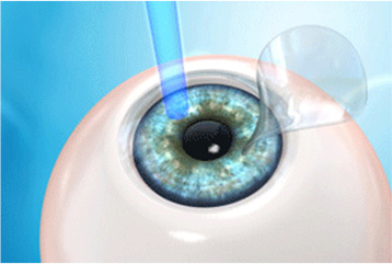
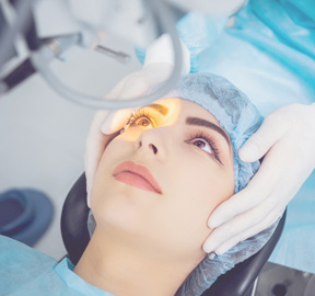
LASER – EXCIMER LASER: For those who do not want to wear glasses or contact lenses, laser surgery is the most indicated, except for rare exceptions
EXCIMER LASER: is the device that will emit the amount and intensity of laser beams in the correct location (ablation) according to the schedule of your surgery. It acts by reshaping the shape of the cornea with a cold laser beam that removes microscopic amounts of tissue (smaller than the thickness of a hair) to create a new curvature of the eye.
Myopia has the cornea more curved, and therefore, the laser flattens. The hypermetrope and the presbyopia have the cornea flatter, and the laser increases the curve. Astigmata has different curves and the laser matches them.
The total surgery time is 5 to 10 minutes each eye. This includes all preparation until finalization of the procedure.
LASER SURGERY STANDARD:
When the degree is only detected with GRENNS, it will be more difficult to obtain the maximum result that this laser can achieve, because it will be manually assigned in the EXCIMER LASER the degree of refraction to be treated. As it varies every 0.25D, patients who have -5.25 D or -5.49 D of myopia, for example, will be treated with the same amount of laser beams. In addition to not treating high-order imperfections, the chances of persisting with some preexisting symptoms such as glare, formation of halos, stars, among others are greater.
CUSTOMIZED LASER SURGERY:
Considered one of the best and most modern surgeries for refraction due to the following advantages: greater comfort, absence of pain, quick recovery and greater safety). The 3D map of the eye, made by theWaveScanWaveFront® aberrometer, will identify all existing imperfections, which will be automatically sent to the EXCIMER LASER, which will be the laser responsible for the final treatment. You will have an exclusive and individualized surgery. It provides a level of precision that meets the specific needs of each eye. The personalized treatment in this method is the one of choice by the best surgeons of the American military.
Recovery is simple and fast. Soon after the surgery, the patient returns to his home and on the next day to routine basic activities. Many patients already report improvement of vision soon after surgery, which is improving throughout the day and over time.
The main goal is to achieve the perfect vision. The result is a daytime and night vision near the ideal (20/20), both far and near. It’s personalized surgery. Made for you, because your eye is unique as the digital pseudedo.
• Clinical Treatment: GREENS and prescription of glasses and contact lenses;
• Surgical Treatment PRK: The cornea is scraped with a blade or similar. 50% laser surgery because one blade and another laser are used.
• Lasik ™ Surgical Treatment: This is also a 50% laser surgery because the (Excimer Laser) is used, but the FLAP is made with a blade that cuts the cornea (microkeratome).
• iLasik ™ Surgery: This is the 100% laser treatment because two laser devices are used for the treatment: Femtosecond to create flap and Excimer to perform the treatment. Diagnostics are performed with the WaveScan WaveFront® aberrometer. It is the safest of the other methods.
Laser refractive surgery is a worldwide consensus among ophthalmologists for bringing great benefits with minimal risk, especially when done 100% laser.
The Visy Institute is distinguished by offering 100% laser refractive eye surgery. Many say that surgery is a laser, but they do not go into detail of the preparation of the cornea for surgery.
This detail is extremely important and decisive in post-surgical recovery. The difference can be between leaving the surgery with a better vision and greater comfort when the preparation of the cornea is done by the IntraLaseiFS® method or living for a week or more postoperative with discomfort (dry eye, irritated, etc.) when using another technique (PRK – scrape the cornea, Microceratome- cut the cornea with blade).
A 100% laser treatment allows the patient to return to daily activities the next day, which does not occur with the PRK and Microkeratome. In addition to providing less risk of infection
100% laser surgery greatly reduces the risks of complications, especially the infection.
See the benefits of the Visy Institute’s All-Laser Laser (Platform – iLasik ™)
Studies in the American Armed Forces after one year of surgery:
- 98% of myopic patients could see 20/20 (perfectly) after treatment;
- 95% were able to pass an habilitation exam, with no need for optical correction;
- 94% reported improvement in quality of life (sports, work, etc.);
- 90% of American Snipers experienced improvement in their abilities after surgery.
- 89% of US Navy pilots assessed significant improvement in the ability to land on Aircraft Carrier after visual laser correction surgery.
Safety:
- Approved FDA, EMEA and ANVISA. (Regulatory agencies of USA, Europe and Brazil.)
- Approximately 32 million Lasik ™ procedures have been performed to date.
- Standard of surgery of pilots of the American Air Force and NASA astronauts.
- Clinical evaluations have shown that the use of IntraLaseiFS® reduces dry eye symptoms by up to 72%.
Preferred of patients:
- 81% of patients choose iLasik ™ with laser corneal preparation rather than traditional blade;
- 75% of the patients chose the postoperative with IntraLaseiFS® when compared to the eye that used the slide;
- Many patients perceive the results immediately after the procedure and the quality of vision continues to improve over the course of days.